Breast Ultrasound Screening Program
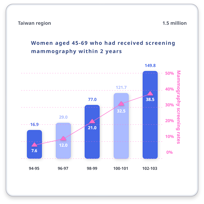
According to Taiwan Cancer Registration Annual Report, breast cancer is the most common cancer among women and with an average of one woman suffering from breast cancer every 36 minutes.

Insufficient Energy
Handheld ultrasound screening has its own evaluation standards in different hospital systems, and the cost of training human resources is high. The technique and experience judgment of the technician or doctor will affect the interpretation of the image.

Loading too High
When doctors write a diagnosis report, they need to view ultrasound images to make a diagnosis. A patient has hundreds of ultrasound images. The large number can easily cause work fatigue and affect judgment.

Patient Loss
The consultation time is already full, leaving no time for patients to do scans. Even if tumors are detected by palpation, ultrasound examinations will take an average of 2 weeks to be arranged, causing patients to be transferred to the hospital for examination.
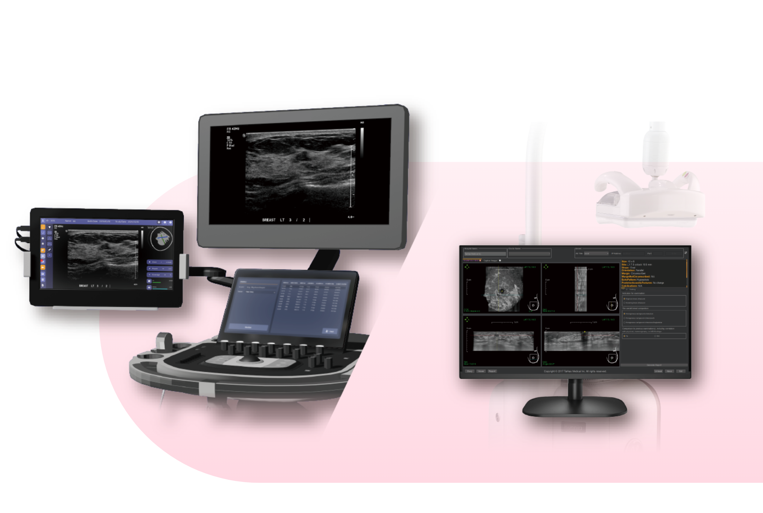
Improve the efficiency of lesion screening
The scanning range is displayed simultaneously during the scan, so that no breasts are missed during scanning and warnings of suspicious lesions are provided in a timely manner, which is equivalent to having an extra specialist nearby to assist in judgment and reduce the missed detection rate.
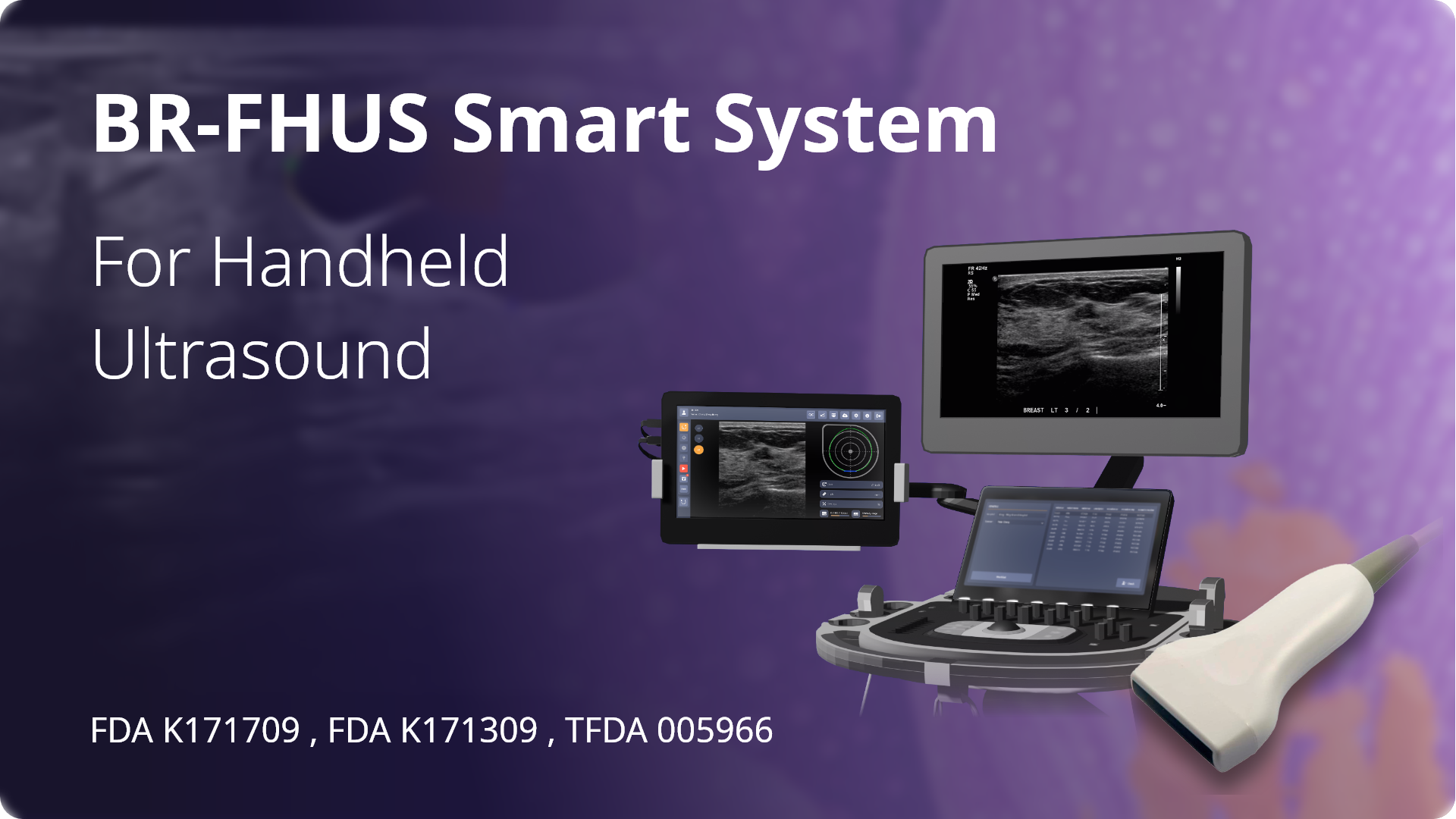
BR-FHUS Smart System
Suitable for experienced radiologists and doctors, AI can assist in prompting suspicious lesions, increase scanning efficiency, and improve examination performance and sensitivity. It is suitable for inexperienced radiologists/other supporting specialists. AI can help to check and remind, improve the detection rate, reduce the missed scan rate, and make the scan more secure.
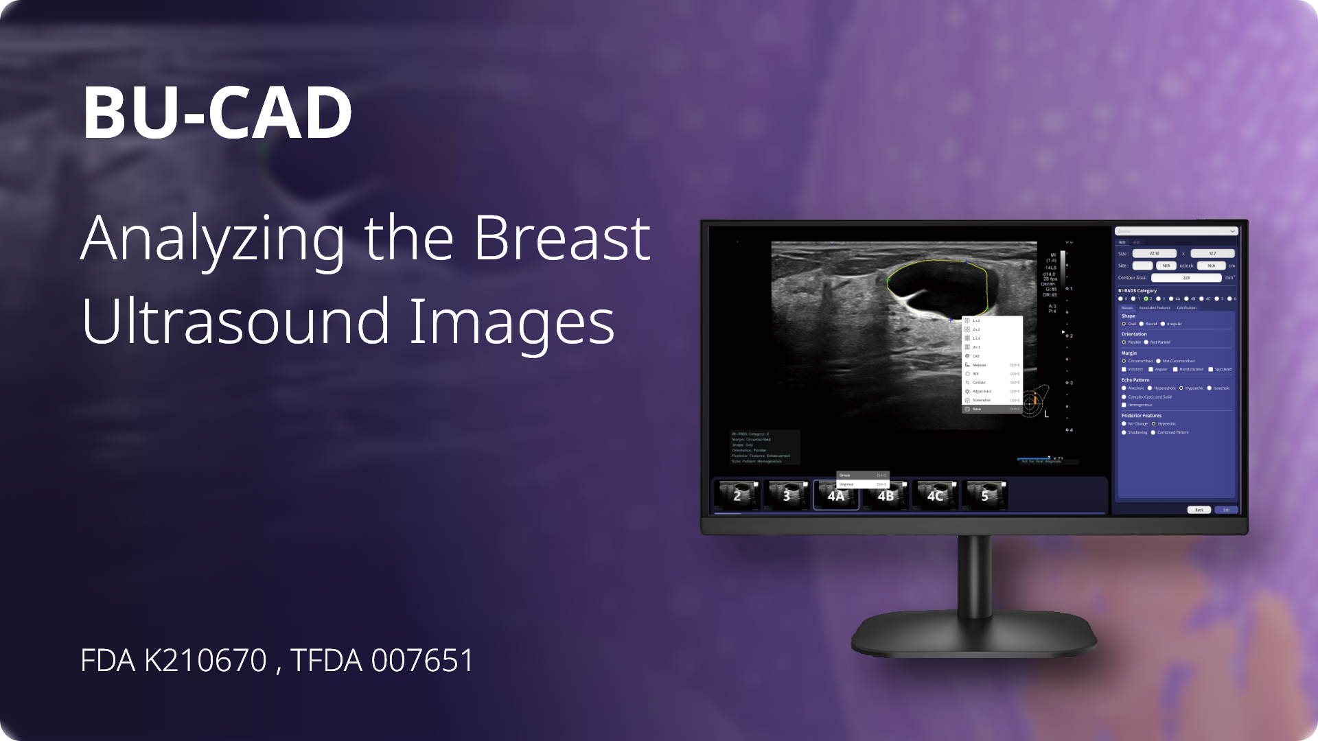
BU-CAD
It is suitable for radiologists, breast surgeons & other specialists who need to interpret breast ultrasound images to improve the efficiency of image reading and quickly produce reports.
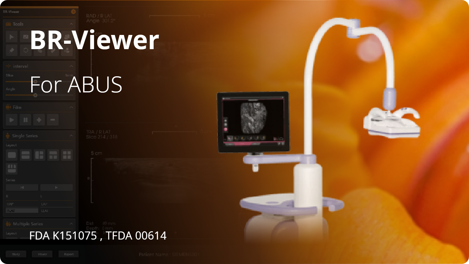
BR-Viewer
ABUS testing produces more images that need to be reviewed, increasing the workload of the reading physician. Using BR-Viewer can improve doctors' reading efficiency, BI-RADS and classify lesions, and quickly generate reports.
-min.gif)